Eyelid
Xanthelasma
Soft yellow cholesterol-rich plaques on the eyelids (xanthelasma palpebrarum) — evaluation of any associated lipid disorder and removal by excision, laser, or chemical treatment.
Eyelid
Soft yellow cholesterol-rich plaques on the eyelids (xanthelasma palpebrarum) — evaluation of any associated lipid disorder and removal by excision, laser, or chemical treatment.
הרופא שלך
Andrew M. Goldbaum, MD
🏅 ASOPRS Fellow
קסנתלסמה — ליתר דיוק xanthelasma palpebrarum — הן פלקים רכים, צהובים, שטוחים או מעט בולטים המורכבים מתאים עמוסים בכולסטרול המצטברים בעור הדק של העפעפיים. הן מופיעות לרוב ליד הפינה הפנימית של העפעף העליון וייתכן שיהיו סימטריות משני הצדדים. קסנתלסמה הן שפירות: הן אינן גידול, אינן זיהומיות ואינן הופכות לסרטן. עבור רבים, הדאגה היא קוסמטית, אך הפלקים יכולים גם להיות אות חיצוני לחילופי חומרים של כולסטרול ולסיכון לבעיות בלב וכלים.
זה מלווה מעמיק להנחיית גידולים ונגעים בעור העפעף הראשית שלנו. קסנתלסמה היא הנפוצה ביותר מבין הקסנתומות של העפעף והיא מוסרת לעתים קרובות יחד עם ניתוח הרמת העפעף.
קסנתלסמה נוצרות כאשר מקרופאגים עשירים בליפידים ("תאי קצף") צוברים עצמם בדרמיס של עור העפעף. בערך חצי מהאנשים הפגועים יש חוסר איזון ליפידי מדיד — רמות LDL כולסטרול מוגבהות, HDL נמוך, או דיסליפידמיה תורשתית — בעוד החצי השני בעל ליפידים בדם נורמליים. ללא קשר לנתוני כולסטרול, מחקרים קושרים קסנתלסמה לסיכון גבוה יותר לטווח ארוך של מחלת לב, לכן הפלקים שווים לקחת ברצינות כאות בריאות. פאנל ליפידים בתחילת הצום ושיחה עם רופא המשפחה שלך הם צעדים הגיוניים ראשונים.
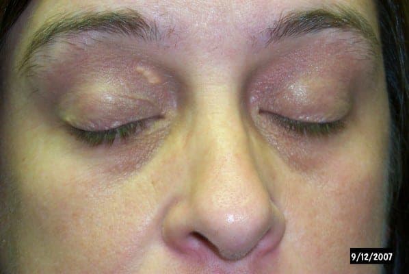
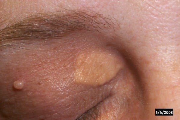
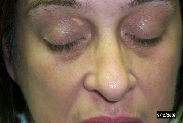
המאפיינים הטיפוסיים הם פלקים צהובים קרימיים, מוגדרים היטב, רכים המרגישים מעט קטיפתיים ישבים ממש מתחת לפני העור. הן גדלות לאט על פני חודשים לשנים, כואבות, וא ל תקרא או מדממות — מאפיינים שעוזרים להבדיל אותן מגידול עפעף אמיתי. כאשר נגע צהוב הוא קשה, לא סדיר, מתעמת או משתנה במהירות, יש להעריך אותו כדי להוציא סרטן עור.
קסנתלסמה היא בדרך כלל אבחנה קלינית — כירורג אופתלמופלסטי יכול לזהות את הפלקים הצהובים האופייניים בבדיקה. מכיוון שקיימים דומים (כולל נגעים שומניים וביל נדיר, קרצינומה שומנית), כל נגע לא אופייני עלול להיות נתון לביופסיה. מעבר לעפעף, ההערכה כוללת פרופיל ליפידים בתחילת הצום וכאשר צוין, בדיקה של סוכרת ותנאים בבלוטת תריס או כבד המשפיעים על ליפידים.
קסנתלסמה אינה דורשת הסרה מטעמי בריאות, אך רבים מהחולים בוחרים בטיפול לשם הופעה. אפשרויות כוללות:
מכיוון שעור העפעף עדין וקרוב לעין, בחירת הטכניקה וטיפול קפדני חשובים; טיפול תוקפני יתר יכול לגרום לצלקות, שינוי פיגמנט, או אי-יישור של העפעף.
קסנתלסמה חוזרות בערך רבע עד 40% מהמקרים, והסיכון גבוה יותר לאחר טיפול חוזר. הישנות היא סבירה יותר כאשר נגעים גדולים, כאשר ארבעת העפעפיים מעורבים וכאשר הפרעה ליפידית בסיסית לא מטופלת. שליטה בכולסטרול ובחירת שיטת ההסרה המתאימה לעובי הנגע שניהם מורידים את הסיכון לחזרה.
שקול הערכה של מומחה אם הפלקים גדלים, מטרידים קוסמטיים, או אם נגע נראה לא טיפוסי (קשה, מתעמת, או משתנה במהירות). כירורג אופתלמופלסטי יכול לאשר את האבחנה, לתאם עבודה ליפידית, ולהסיר את הפלקים עם תשומת לב לתפקוד וחזות של העפעף.
ראה כירורג אופתלמופלסטי
נגעי העפעף יושבים במילימטרים מהעין, שם הסרה והשחזור דורשים טיפול של מומחה. מצא כירורג אופתלמופלסטי מאומן ASOPRS ליד ביתך.