Eyelid
Surgical correction of congenital eyelid and orbital conditions present from birth — ptosis, blocked tear ducts, hemangiomas, and more.
Medically reviewed by Andrew M. Goldbaum, MDOculoplastic SurgeonLast updated June 2026
Congenital conditions of the eyelid, orbit, and lacrimal system are present from birth or become apparent in early childhood. They range from minor cosmetic variations to conditions that threaten vision through amblyopia — the brain’s suppression of input from an eye whose image is chronically blurred, blocked, or misaligned. Early detection and appropriately timed surgery are critical for visual development.
ASOPRS-trained oculoplastic surgeons care for children with eyelid and orbital conditions that require surgical expertise, coordinating with pediatric ophthalmology for vision assessment and amblyopia management. The sections below describe the most common congenital conditions seen in this practice.
Congenital ptosis is a drooping of the upper eyelid present from birth, most commonly due to a developmental dysgenesis of the levator palpebrae superioris muscle. Unlike acquired ptosis in adults, in which the muscle is normal but its tendon is stretched, congenital ptosis is caused by fibrofatty replacement of levator muscle fibers — producing a weak, non-elastic muscle that neither lifts well nor relaxes fully.
The visual system develops in the first 7–10 years of life. During this critical period, both eyes must deliver a clear, focused image to the brain for normal visual development. Ptosis threatens vision in two ways:
Children also compensate for ptosis by adopting a chin-up head posture or by raising the brow. These compensatory postures do not protect vision but indicate that the child is attempting to see under a drooped lid — a sign that the ptosis is visually significant.
The surgical approach depends on the degree of levator function (how many millimeters the lid moves from down-gaze to up-gaze):
Full details of ptosis evaluation and surgical technique are on the Ptosis page.
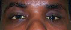
Congenital nasolacrimal duct obstruction is the most common cause of tearing and eye discharge in infants. Approximately 6% of newborns have an imperforate membrane at the distal nasolacrimal duct (the valve of Hasner) that normally opens at or shortly after birth. Tears that cannot drain stagnate and frequently become infected.
CNLDO presents in one of four ways:
The vast majority of simple CNLDO resolves spontaneously: 90% open by 12 months of age. The accepted initial management is Crigler massage — digital compression over the lacrimal sac 5–10 times, 2–3 times daily, to create a hydrostatic pressure wave that opens the distal membrane. Topical antibiotics are used episodically for infectious exacerbations but do not cure the obstruction.
If spontaneous resolution has not occurred by 9–12 months, probing is offered:
The orbit develops from a complex interaction of neural crest cells, mesoderm, and optic vesicle signals. Disruption of these processes can result in abnormalities of orbital size, shape, and contents.
Microphthalmos (abnormally small eye) and anophthalmos (absent eye) represent a spectrum of severe developmental failures of the optic vesicle. They may be isolated or part of a systemic syndrome. Anophthalmos requires early intervention with conformers and orbital expanders to stimulate orbital growth — the orbital bones depend on the eye’s presence to grow normally. Without treatment, the orbit remains small and socket reconstruction in adulthood is challenging.
Dermoid cysts are the most common benign orbital mass of childhood — choristomas that arise at embryonic suture lines from trapped ectoderm. The superolateral orbital rim (frontozygomatic suture) is the classic location. They are smooth, non-tender, freely mobile masses that do not transilluminate. Most present in the first decade. Management is complete surgical excision — the cyst wall must be intact; rupture causes severe granulomatous inflammation. Deep dermoids extending intracranially require CT planning before surgery.
The most common orbital tumor of infancy. Periorbital infantile hemangiomas may extend into the orbit, producing ptosis and proptosis. They proliferate rapidly in the first year of life, then involute slowly over years. Treatment with oral propranolol (1–3 mg/kg/day) is now first-line for lesions that threaten vision or cause significant deformity.
Blepharophimosis, ptosis, and epicanthus inversus syndrome (BPES) is an autosomal dominant condition caused by mutations in the FOXL2 gene. The defining features are:
Surgical correction is staged: medial canthal tendon plication (Y-V or W-plasty) is performed at 12–18 months to widen the fissure and correct telecanthus before ptosis repair. Ptosis repair with frontalis sling typically follows. BPES Type I is associated with premature ovarian failure; genetic counseling and gynecologic follow-up are indicated for affected females.
Epiblepharon is a horizontal fold of skin just below the lower lid margin that redirects lashes toward the cornea. It is particularly common in East Asian children. Most cases resolve spontaneously by age 5 as the face elongates and orbicularis atrophies. Surgery (excision of a skin-muscle strip) is indicated for persistent corneal staining, pain, or visual disturbance.
Congenital entropion is an inward rotation of the lid margin itself. It is less common than epiblepharon and is more likely to require surgical correction. Corneal staining confirms lash abrasion and indicates the need for treatment.
A coloboma is a notch or cleft in the eyelid present from birth, usually affecting the upper lid at the junction of the medial and central thirds. Small colobomas may not cause corneal exposure; large defects require early reconstruction to protect the cornea from exposure keratopathy. Eyelid colobomas may be isolated or associated with Goldenhar syndrome (oculo-auriculo-vertebral spectrum), which includes ear and vertebral anomalies.
Distichiasis is an accessory row of eyelashes arising from the meibomian gland orifices, directed toward the cornea. The lashes are typically fine and soft; many patients are asymptomatic. Symptomatic cases with corneal irritation or staining are treated with cryotherapy to the posterior lamella (freeze-thaw cycles to ablate aberrant follicles), electrolysis, or lid-splitting procedures for localized areas.
Schedule a consultation with Andrew M. Goldbaum, MD to learn if this procedure is right for you.