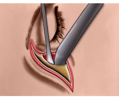
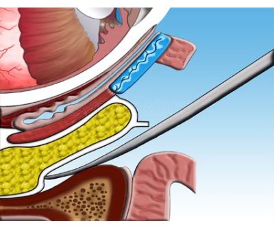
In order to create additional space on the outer orbit, the surgeon may remove some of this bone with (as seen here) or thin the bone in this area.
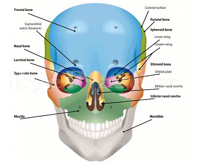
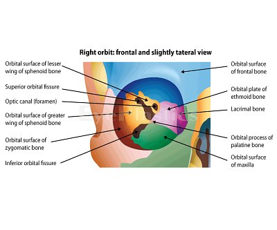
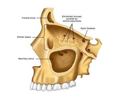
In humans, seven bones make up the bony orbit: Frontal bone (Pars orbitalis), Lacrimal bone, Ethmoid bone (Lamina papyracea), Zygomatic bone (Orbital process of the zygomatic bone), Maxillary bone (Orbital surface of the body of the maxilla), Palatine bone (Orbital process of palatine bone), Sphenoid bone (Greater and lesser wings).
Foramina and openings include: Optical foramen, Superior orbital fissure, Inferior orbital fissure, Anterior ethmoidal foramen, Posterior ethmoidal foramen, Infraorbital foramen, Supraorbital foramen, Naso-lacrimal canal opening, Zygomatic orbital foramen.
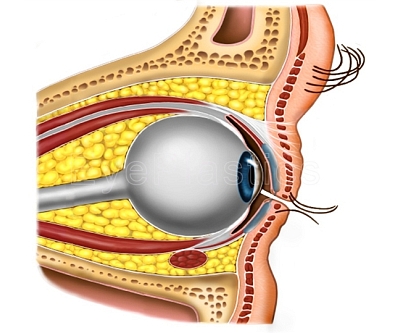
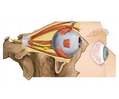
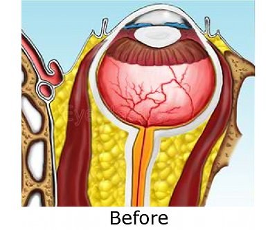
In the orbit, fat tissue, which surrounds the eyeball and its muscles, keeps the rotation smooth with respect to the center of the eye. If excess liquid is collected in the fat cushion tissue, the eye may protrude (known also as exophthalmos).
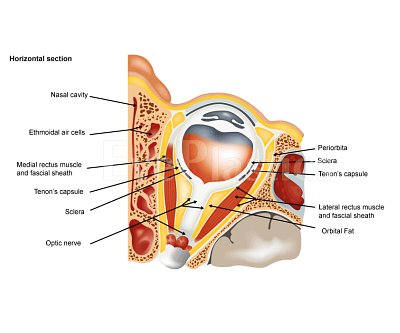
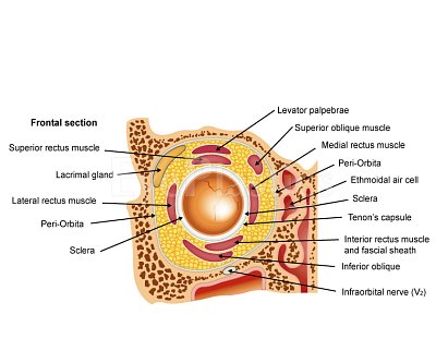
Cross section through the orbit illustrating the relative position of the eyeball, extraocular muscles and other anatomical structures.
In anatomy, a nasal concha (or turbinate) is a long, narrow and curled bone shelf (shaped like an elongated sea-shell) which protrudes into the breathing passage of the nose. In humans, the turbinates divide the nasal airway into four groove-like air passages.
In anatomy, a nasal concha (or turbinate) is a long, narrow and curled bone shelf (shaped like an elongated sea-shell) which protrudes into the breathing passage of the nose. In humans, the turbinates divide the nasal airway into four groove-like air passages (Alternative view).
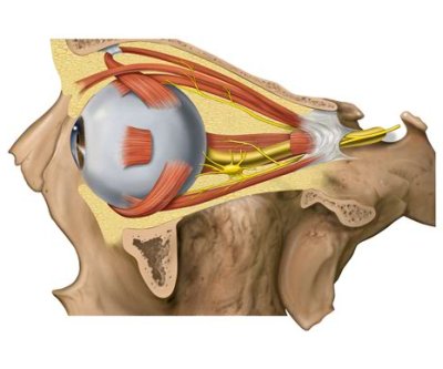
Axial (top-down) view of the orbit, illustrating the eyeball, muscles and nerves.
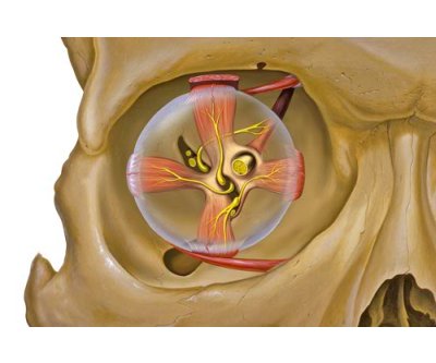
Front view of the orbit, illustrating the eyeball, muscles and nerves.
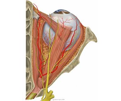
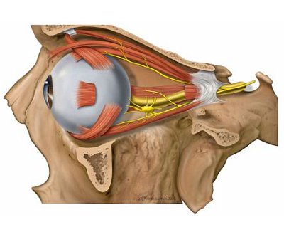
Side (Sagittal) view of the orbit, illustrating the eyeball, muscles and nerves, with orbital fat.
Side view (sagittal) of the orbit, illustrating the eyeball, muscles and nerves, WITHOUT orbital fat.
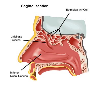
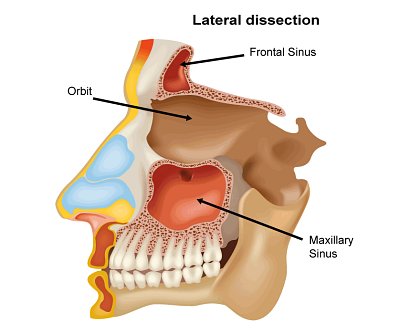
Paranasal sinuses are a grouop of four paired air-filled spaces, that surround the nasal cavity (maxillary sinuses), above and between the eyes (ethmoid sinuses and frontal sinuses), and behind the ethmoids (sphenoid sinuses). The sinuses are named for the facial bones behind which they are located.
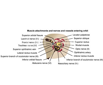
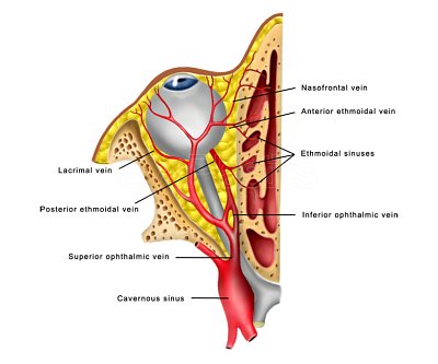
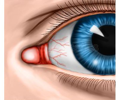
Orbital Contents include: Eyeball, Fascias (Orbital, Bulbar) Extraocular muscles (Levator Palpebrae Superioris, Superior, Inferior, Lateral and Medial Rectus muscles, Superior and Inferior Oblique Muscles), Nerves (cranial nerves II, III, IV, V and VI), Blood vessels, Extraocular Fat, Lacrimal gland, Lacrimal sac, Nasolacrimal duct, Eyelids.